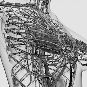
Women's Health and Infertility
Our interventional care for women includes:
-
Uterine Fibroid
Embolization -
Pelvic Congestion
Syndrome - Breast Biopsy
University Radiology Interventional Radiology
- Oncology
- Organ Biopsy (Liver, Thyroid, Lymph Node)
- Brain and Spine Interventions
- Bone, Muscle and Joint Pain
- Liver and Gallbladder Disease
- Women's Health and Infertility
- Men's Health and Infertility
- Kidney and Urinary Disease
- Cyst and Fluid Drainage
- Arterial Disease
- Deep Venous Disease
- Vascular Access and Dialysis
-
Interventional Radiology
Physicians - Advanced Practice Providers
Women's Health and Infertility
University Radiology's interventional radiologists are experts in providing advanced diagnostic imaging and specialized, minimally invasive treatments for many common health conditions in women. By taking advantage of sophisticated medical imaging, our radiologists are treating painful and chronic conditions such as uterine fibroids without surgery. For patients, that means less pain and a shorter recovery compared with traditional surgery. Some of the treatments we provide include:
Uterine Fibroid Embolization
Uterine fibroids are benign growths that arise from the muscular wall of the uterus. It is estimated that up to 40 percent of women over the age of 35 have fibroids, which may vary in size from less than an inch to the size of a 5-month pregnancy. During uterine fibroid embolization, small particles are injected into the arteries that supply blood to the fibroids. These particles block the blood supply to the fibroids, causing them to shrink.
University Radiology was among the first groups in New Jersey to perform uterine fibroid embolization in 1996, and we have performed more than 1,500 procedures.
What are the benefits?
- Resolves symptoms caused by uterine fibroids, including heavy, painful, prolonged monthly periods, anemia, fatigue, frequent urination and pain or pressure in the hips and back.
- A minimally invasive alternative to traditional open surgery to remove the uterus (hysterectomy), or removal of the fibroid (myomectomy), with a faster recovery time and a shorter hospital stay.
- Preserves the uterus.
How does the procedure work?
After numbing the skin at the hip region, your interventional radiologist will make a tiny skin nick, no larger than one-half the width of a fingernail. A small tube (catheter) is inserted into an artery in the hip and is guided into one of the uterine arteries under X-ray guidance. Tiny particles are injected into the uterine artery, which stops or slows the blood flow to the uterus and to the fibroids. The procedure is then repeated for the uterine artery on the opposite side of the uterus. The procedure takes about 1 to 2 hours and is performed under moderate sedation. You will likely spend one night in the hospital before returning home the following morning. We expect that you will resume light activities in a few days and return to normal activities 7 to 10 days after the procedure.
Pelvic Congestion Syndrome
Chronic pelvic pain can be very difficult to diagnose and treat because it has many different and often overlapping causes. Recent advances have shown the pain may be due to ovarian/pelvic varicose veins - dilations of normal veins that allow blood to pool. The veins compensate by getting bigger, which can cause pain and aches in the lower belly, pelvic region and lower back. At University Radiology, we offer a procedure called ovarian/pelvic vein embolization, which blocks the flow of blood in faulty, enlarged veins that are causing pain. The body naturally re-directs blood to other healthy pathways.
What are the benefits?
- A minimally invasive treatment for the ovarian/pelvic varicose veins, which may lead to relief of chronic pelvic pain.
- Reduction in size of the varicose veins in the pelvis, genitals and thigh.
- Less invasive than other options such as surgical ligation of the ovarian veins or hysterectomy and salpingo-oophorectomy.
How does the procedure work?
After numbing the skin at the hip region, your interventional radiologist will make a tiny skin nick, no larger than one-half the width of a fingernail. A small tube (catheter) is inserted into a vein in the groin and is guided into an ovarian vein under X-ray guidance. Dye will be injected to confirm that there is backing up of blood in the ovarian and pelvic veins. If there is significant backup of blood in the veins, an embolization will be performed. By using coils, a vein occluding liquid/foam or both, your interventional radiologist will block the blood flow in the problematic veins that are causing pain. The embolization is usually performed on both ovarian veins during the same procedure. The procedure takes about 1 to 2 hours and you may spend a night in the hospital. We expect that you will resume normal activities in a few days after the procedure.
Breast Biopsy
When a physical exam or a medical imaging test such as mammography finds a breast change or abnormality, your doctor may order a breast biopsy. During a breast biopsy, a tissue sample is taken from the suspicious area of the breast for testing. Fortunately, most breast changes are not cancer. However, a biopsy is the only way for your doctor to know for sure.
What are the benefits?
- A less invasive method than surgical biopsy to obtain breast tissue samples for testing.
- Leaves little or no scarring and can be performed in less than an hour.
How does the procedure work?
Breast biopsies are taken using a needle guided by imaging - a mammogram, breast MRI scan or ultrasound. The medical images show your specially trained breast radiologist exactly where to place the needle and take the sample. At University Radiology, we offer three types of breast biopsy:
- Fine needle aspiration: Your radiologist uses a very thin, hollow needle attached to a syringe to draw out a small amount of tissue from the area of concern. The needle is thinner than the one used for blood tests.
- Core needle: Your radiologist uses a large hollow needle to remove a small core of issue (about one-eighth inch in diameter and one-half inch long) from the area of concern. Usually, several cores are removed.
- Vacuum-assisted: Your radiologist uses a suction device that pulls tissue into a hollow needle. More tissue can be removed with this type of biopsy than with a core needle biopsy. Usually, the radiologist removes several samples.
University Radiology
Interventional Radiology
- Oncology
-
Organ Biopsy
(Liver, Thyroid, Lymph Node) - Brain and Spine Interventions
- Bone, Muscle and Joint Pain
- Liver and Gallbladder Disease
- Women's Health and Infertility
- Men's Health and Infertility
- Kidney and Urinary Disease
- Cyst and Fluid Drainage
- Arterial Disease
- Deep Venous Disease
- Vascular Access and Dialysis
-
Interventional Radiology
Physicians - Advanced Practice Providers