Diagnostic X-ray
- PATIENT PREPARATION
- Overview
- Arthrogram
- Barium Enema
- Cystogram
- DXA
- Esophagram
- Hysterosalpingogram
- IVP (Intravenous Pyelogram or Intravenous Urogram)
- Mammography - Digital Mammogram
- Myelogram
- Small Bowel Series
- Upper GI Series
- UGI and Small Bowel
- VCUG (Voiding Cystourethrogram)
- X-ray Studies of the Head, Chest, Spine, Abdomen, or Extremities
Diagnostic X-ray Overview
Overview
Diagnostic x-ray procedures are studies that use ionizing radiation to produce an image on x-ray film. Diagnostic x-ray studies can be done on all body parts.
Contrast media are used in various x-ray procedures to enhance the image. The x-ray films are interpreted by the radiologist and the report and/or films are sent to the referring physician.
Each type of diagnostic x-ray procedure is described briefly below. If patient preparation is required, the patient should follow prep instructions listed on the Adult or Pediatric Prep Sheet Instructions form available through our offices or this site.
All diagnostic x-ray exams require a written prescription or referral stating which exam is being ordered and the patient’s signs and symptoms indicating medical necessity of the procedure.
Patients must be screened for asthma, allergies and diabetes and any use of an inhaler prior to exams requiring contrast. If the patient is diabetic, it is critical that we know what medications they are taking.
Patients should bring previous films or related exams to their appointment. This information is very important and will expedite the report of the diagnosis back to the referring physician.
Common X-ray Studies & Patient Preparation
Arthrogram
X-ray study performed for the visualization of cartilage, ligaments and any loose bodies in a joint. The study requires an injection of contrast or contrast and air into the joint space. There is no patient preparation for this exam.
Barium Enema
X-ray study of the large intestine using barium and air introduced through the rectum. Patient prep is required.
Cystogram
X-ray study of the bladder during which contrast material is dripped into the bladder via a catheter. There is no preparation for this exam.
DXA
X-ray study of the bones (usually the spine and hips) to check for osteoporosis (bone loss). There is no preparation for this exam. See also Screening for Health.
Esophagram
X-ray study of the esophagus which requires the patient to drink a barium solution. Patient prep is required.
Hysterosalpingogram
X-ray study of the uterus and fallopian tubes during which contrast is injected through the cervical opening. Patient prep is required.
IVP (Intravenous Pyelogram or Intravenous Urogram)
X-ray study of the kidneys and collecting system in which contrast is injected into a vein. Patient prep is required.
Mammography - Digital Mammogram
X-ray study of the breast that uses a low level of radiation. Mammography can detect lesions in breast tissue which might otherwise go unnoticed because they are too small to be felt. This makes mammography particularly valuable as a screening tool in detecting early breast cancer.
There may be discomfort associated with a mammogram due to the compression which is necessary to obtain a clear picture of the internal structures. The compression is not dangerous and does not damage breast tissue. It improves the accuracy of the mammogram and also reduces the amount of radiation needed for the test. The discomfort is temporary and can be relieved by oral analgesics.
Routine mammograms should be scheduled within the first 14 days after the patient's period to reduce the amount of potential discomfort from the compression.
Routine mammograms are interpreted by Mammography Specialists at the University Radiology Digital Screening Mammography Program. Two views of each breast are taken at the time of the office visit. If additional views are needed to make the diagnosis, the patient is contacted to return for specialized spot or magnification views as requested by the Radiologist. If no additional views are needed, a report is sent to the patient and the referring physician.
Please note that women under 30 years old are only scheduled for a mammogram if there is an abnormality or severe family history.
The patient is requested not to use any powders, creams or deodorant under the arms or around the breast area in preparation for the exam. Patients are asked to please bring any prior mammogram studies from other facilities with them for their appointment. Please note that it is very important that all mammograms be returned to us so that they can be used in the future for comparison. As part of the complete exam, current films are always compared with prior studies. See also Women's Diagnostics
Myelogram
X-ray study of the spinal column and nerves. Under fluoroscopic guidance, the radiologist will place a needle into the spine ("spinal tap"). Through this same needle, contrast is injected and subsequent x-rays obtained. A nurse from University Radiology will contact the patient after the appointment is scheduled to review any necessary preparation. It is critical, therefore, that we have the home and work phone numbers of the patient.
Small Bowel Series
X-ray study of the small intestine which requires the patient to drink a barium solution. Patient prep is required.
Upper GI Series
X-ray study of the esophagus, stomach and small intestine which requires the patient to drink a barium solution with gas granules. Patient prep is required.
UGI and Small Bowel
X-ray study of the esophagus, stomach and small intestine which requires the patient to drink a barium solution with gas granules. Patient prep is required.
VCUG (Voiding Cystourethrogram)
X-ray study of the bladder and urethra. Contrast is placed into the bladder via a catheter. After filling the bladder, the catheter is removed and pictures are taken as the patient voids. There is no preparation for this exam.
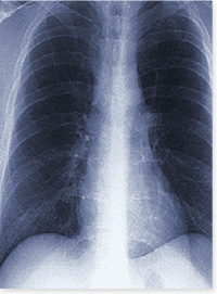
X-ray Studies of the Head, Chest, Spine, Abdomen, or Extremities
No patient preparation is required. Please send any prior studies for comparison.

- PATIENT PREPARATION
- Overview
- Arthrogram
- Barium Enema
- Cystogram
- DXA
- Esophagram
- Hysterosalpingogram
- IVP (Intravenous Pyelogram or Intravenous Urogram)
- Mammography - Digital Mammogram
- Myelogram
- Small Bowel Series
- Upper GI Series
- UGI and Small Bowel
- VCUG (Voiding Cystourethrogram)
- X-ray Studies of the Head, Chest, Spine, Abdomen, or Extremities