Nuclear Medicine
- Overview
- The Procedure
- Bone Scan
- Cardiac Scan
- Gallium Scan
- Gastric Emptying Scan (Liquid and Solid)
- Gastroesophageal Reflux Scan
- Hepatobiliary Scan
- Lymphoscintigraphy
- Meckel's Scan
- Nuclear Cystogram
- Parathyroid Scan
- Renal Flow
- SPECT Scan
- Thyroid Uptake and Scan
- Whole Body Iodine Uptake Scan
- Nuclear Medicine Therapy
Nuclear Medicine Overview
Overview
Nuclear Medicine is the medical specialty that uses radioactive tracers to aid in the diagnosis and treatment of a wide variety of diseases. After the tracer is administered, a gamma camera and computer are used to create images or scans of important body and organ functions. The scans are usually performed in conjunction with other imaging studies which give more anatomic information of the regions studied.
Nuclear medicine studies use radiation. The dose to the patient varies depending on the test, but the range is similar to x-rays and CT scans. Children receive a lower amount of radiation that is based on their weight. Usually, no special precautions need to be taken after the study and normal activity can be resumed immediately.
In general, the actual tracers cause no pain or any other symptom. University Radiology has extensive experience using these tracers; the physicians and technologists are trained and certified in their use; and reactions to the tracer are very rare.
Patient preparation for nuclear medicine studies is highly variable and a nuclear medicine technologist or nurse will contact the patient prior to the examination if any special preparation is required.
A brief description of each Nuclear Medicine is shown below.
Note Regarding Pregnancy Testing: As with all tests involving radiation, patients will be asked whether or not they may be pregnant. Most nuclear medicine scans are avoided or postponed if there is a possibility of pregnancy in order to avoid risk to the fetus.
Even though the patient may be certain that she is not pregnant, University Radiology protocols may require a blood pregnancy test. Be assured that this requirement is applied uniformly, the only goal is to guarantee patient safety, and nuclear medicine protocols do not allow exceptions to the rule.
The Procedure
- The nurse and/or technologist will review the procedure with the patient and answer all questions. Also, the patient will be asked relevant questions regarding their medical history. This helps the nuclear medicine physician interpret the study.
- A radioactive agent is administered either intravenously or by mouth.
- A gamma camera/computer is used to create an image of radioactivity in different parts of the body, as required by the nature of the study. The scan is recorded in a computer and on photographic film.
- The duration of the test varies depending on the specific test ordered by the physician. Some tests take less than an hour to complete, while other tests may take up to several hours (see below).
- A nuclear medicine physician interprets the results, and sends a report to the referring physician.
All Nuclear Medicine studies require a written prescription or referral stating the exam being ordered and the patient’s signs and symptoms indicating medical necessity of the procedure. If we do not have the written prescription in hand, the radioisotope cannot be administered.
Frequently Ordered Nuclear Medicine Studies
Bone Scan
Used for early detection of fractures, tumors, infection and arthritis.
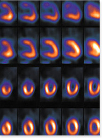
Cardiac Scan
Muga scan is used to evaluate the function of the heart as a pump. Myocardial perfusion is used to evaluate the adequacy of the coronary blood flow to the heart muscle.
Gallium Scan
Detects soft tissue and skeletal infections and tumors.
Gastric Emptying Scan (Liquid and Solid)
Evaluates how well your stomach empties.
Gastroesophageal Reflux Scan
Evaluates gastroesophageal reflux (usually in children).
Hepatobiliary Scan
Detects acute cholecystitis. May also be used to evaluate for chronic gallbladder abnormalities.
Lymphoscintigraphy
Delineates the path of lymph channels prior to surgical removal of malignant tumors.
Meckel's Scan
Detects bleeding from a Meckel's diverticulum.
Nuclear Cystogram
Detects reflux from urinary bladder to ureters (usually in children).
Parathyroid Scan
Detects presence of parathyroid adenoma or hyperplasia in patients with abnormally high serum calcium levels.
Renal Scan
Evaluates kidney blood flow, urine formation and urinary obstruction.
SPECT Scan
Computed tomographic radionuclide study to improve detection of small lesions, usually as a part of a study using planar imaging.
Thyroid Uptake and Scan
Evaluates thyroid nodules and how well the thyroid is functioning.
Whole Body Iodine Uptake Scan
Detects remnants of functioning thyroid and thyroid cancer after surgery for thyroid cancer.
Nuclear Medicine Therapy
- Overview
- The Procedure
- Bone Scan
- Cardiac Scan
- Gallium Scan
- Gastric Emptying Scan (Liquid and Solid)
- Gastroesophageal Reflux Scan
- Hepatobiliary Scan
- Lymphoscintigraphy
- Meckel's Scan
- Nuclear Cystogram
- Parathyroid Scan
- Renal Flow
- SPECT Scan
- Thyroid Uptake and Scan
- Whole Body Iodine Uptake Scan
- Nuclear Medicine Therapy